Home
Uncategories
Anatomy Of Ribs And Sternum / Bones And Joints In The Thoracic Region Dummies / The sternum is a flat, long bone that forms the medial and anterior part of the thoracic cage.
Anatomy Of Ribs And Sternum / Bones And Joints In The Thoracic Region Dummies / The sternum is a flat, long bone that forms the medial and anterior part of the thoracic cage.
Anatomy Of Ribs And Sternum / Bones And Joints In The Thoracic Region Dummies / The sternum is a flat, long bone that forms the medial and anterior part of the thoracic cage.. It consists of the ribs, the sternum, and the thoracic vertebrae, to which the ribs articulate. The sternum, commonly known as the breastbone, is a long, narrow flat bone that serves as the keystone of the rib cage and stabilizes the thoracic skeleton. The classification of human ribs. An exception to this rule is that the first rib articulates with the first thoracic vertebra only. Diagnose and treat somatic dysfunctions of the clavicle, sternum, and ribs.
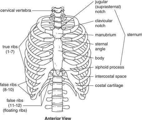
Anterior view of the thoracic cage image source. This guide gives a general overview of the anatomy of the thoracic spine. There are twelve pairs of ribs. It has clear front, side, and back planes. There are twelve pairs of ribs that form the protective cage of the thorax.
Sternum With Rib Cartilage Anatomical Models Anatomical Charts from cdn3.volusion.com There is a printable worksheet available for download here so you can take the quiz with pen and paper. Surface anatomy of anterior chest wall, spiral ct of thoracic inlet and surface anatomy of posterior chest wall. It is an atypical rib and is an important anatomical landmark and is one of the borders of the superior thoracic aperture. An exception to this rule is that the first rib articulates with the first thoracic vertebra only. Important clinical anatomy of the head, neck, and back. The final two pairs of ribs are floating ribs and the cartilage of these ribs tends to end within the abdominal musculature. Sternum, costal cartilages, and ribs image source: Learn about sternum thorax ribs anatomy with free interactive flashcards.
Join the sternum and clavicles.
Sternum is a part of the skeletal system. The thoracic cage (rib cage) is the skeletal framework of the thoracic wall, which encloses the thoracic cavity. Moore'sanatomy and osteology human anatomy vertebra bones of the upper limb how to classified ribs and their shapes and fully describe human anatomy of upper parts. Learn more about the skeletal system with quizzes and labelling exercises. Join us in this video where we show the sternum and rib articulation anatomy through the use of a model. Sternum, costal cartilages, and ribs image source: There are twelve pairs of ribs. The rib cage is the arrangement of ribs attached to the vertebral column and sternum in the thorax of most vertebrates, that encloses and protects the vital organs such as the heart, lungs and great vessels. It also includes some facts regarding pathophysiology in this region. Important clinical anatomy of the head, neck, and back. The body is the middle part of the sternum and is also the longest. Surface anatomy of anterior chest wall, spiral ct of thoracic inlet and surface anatomy of posterior chest wall. Each pair articulates with a.
Ossification.—the sternum originally consists of two cartilaginous bars, situated one on either side of the median plane and connected with the cartilages of the upper nine ribs of its. This often has little impact on function or treatment following injury but can however, cartilaginous connectors between the sternum and each of the upper six ribs assist with minor motions that occur with each breath. The classification of human ribs. There are twelve pairs of ribs. Sternum, costal cartilages, and ribs image source:
Sternum And Ribs Anatomy Anatomy Drawing Diagram from lh3.googleusercontent.com Each pair articulates with a. There is a printable worksheet available for download here so you can take the quiz with pen and paper. Try to be as accurate as you can with them. Surface anatomy and surface markings bibliographic record list of illustrations subject index. The chest wall is formed from the sternum anteriorly, 12 pairs of ribs, costal cartilages and intercostal muscles laterally, and the thoracic vertebrae posteriorly. The sternum, commonly known as the breastbone, is a long, narrow flat bone that serves as the keystone of the rib cage and stabilizes the thoracic skeleton. Manubrium, body (gladiolus), and xiphoid process 1. It connects to the ribs via cartilage and forms the front of the rib cage, thus helping to protect the heart, lungs, and major blood vessels from injury.
Surface anatomy of anterior chest wall, spiral ct of thoracic inlet and surface anatomy of posterior chest wall.
It discusses the specific anatomy of the ribs and costal cartilages, along with the sternum. The ribs stretches posteriorly from thoracic vertebrae to the anterior lateral edges of the sternum. It lies on the anterior thoracic wall in the middle. It is an atypical rib and is an important anatomical landmark and is one of the borders of the superior thoracic aperture. Ribs are greatly reduced and never meet sternum each thoracic rib has two segments:. Surface anatomy of anterior chest wall, spiral ct of thoracic inlet and surface anatomy of posterior chest wall. Manubrium, body (gladiolus), and xiphoid process 1. Coastal cartilages are joined to the. The sternum is a flat, long bone that forms the medial and anterior part of the thoracic cage. An exception to this rule is that the first rib articulates with the first thoracic vertebra only. Individual ribs have a bony dorsal part, a body of rib, and ventral costal cartilage. This is an online quiz called ribs and sternum anatomy. Learn about sternum thorax ribs anatomy with free interactive flashcards.
The thoracic cage consists of the 12 thoracic vertebrae, the associated intervertebral discs, 12 pairs of ribs with their costal cartilages, and the sternum. There is a printable worksheet available for download here so you can take the quiz with pen and paper. Anatomical variations of the sternum include varying sizes of the sternal angle. Ossification.—the sternum originally consists of two cartilaginous bars, situated one on either side of the median plane and connected with the cartilages of the upper nine ribs of its. Important clinical anatomy of the head, neck, and back.
The Thoracic Cage Scientist Cindy from www.scientistcindy.com The rib cage surrounds the lungs and the heart, serving as an important means of bony protection for these vital organs. It has clear front, side, and back planes. Regional vertebrae (cervical, thoracic, lumbar), rib, sternum, os coxae, clavicle, scapula, humerus, ulna and radius for dr. Describe the bony and cartilaginous articulations of the sternum and clavicle. It also includes some facts regarding pathophysiology in this region. Moore'sanatomy and osteology human anatomy vertebra bones of the upper limb how to classified ribs and their shapes and fully describe human anatomy of upper parts. Ossification.—the sternum originally consists of two cartilaginous bars, situated one on either side of the median plane and connected with the cartilages of the upper nine ribs of its. There is a printable worksheet available for download here so you can take the quiz with pen and paper.
It also includes some facts regarding pathophysiology in this region.
Sternum is a part of the skeletal system. Construct a robo skelly rib cage and the pelvis using the bucket method. Your third through seventh set of ribs are connected to the body of the sternum via cartilage. Manubrium, body (gladiolus), and xiphoid process 1. The first portion, the manubrium, articulates with both the clavicle and first rib and is therefore. The sternum is comprised of three distinctive portions: Learn all about this bone using our interactive anatomy image and detailed descriptions of its parts and function! It is an atypical rib and is an important anatomical landmark and is one of the borders of the superior thoracic aperture. The chest wall is formed from the sternum anteriorly, 12 pairs of ribs, costal cartilages and intercostal muscles laterally, and the thoracic vertebrae posteriorly. Ribs are greatly reduced and never meet sternum each thoracic rib has two segments:. Describe the bony and cartilaginous articulations of the sternum and clavicle. The body is the middle part of the sternum and is also the longest. Diagnose and treat somatic dysfunctions of the clavicle, sternum, and ribs.
There are twelve pairs of ribs that form the protective cage of the thorax anatomy of ribs. There are two classifications of ribs.


0 Comments:
Post a Comment